The hand anatomy mini-series – Muscular Layers
If you haven’t already read or would like a recap of our hand anatomy blogs, please follow the links below to update your understanding of the anatomy of the hand –
Bony Anatomy –

Connective Tissue Anatomy –

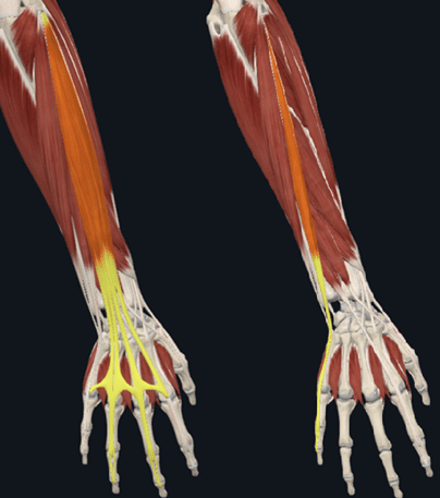
Figure 1 shows the extensor digitorum insertion onto the middle and distal phalanges of the second to fifth digits of the hand on the left. The right image shows extensor digiti minimi inserting onto the extensor expansion of the fifth digit. Both muscles originate via the common extensor tendon from the lateral epicondyle of the humerus and extend the hand and digits of their insertions.
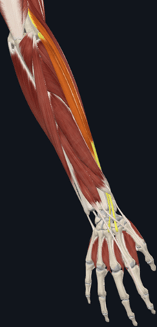
Continuing with our other wrist extensors, Figure 2 shows the extensor carpi radialus longus and brevis. ECRL is shown with a more superior origin from the supracondylar ridge of the humerus, and the ECRB originating more distally via the aforementioned common extensor tendon. Again, these muscles extend and abduct the hand, also termed radial deviation.
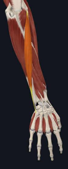
Figure 3 shows the final of the four muscles originating from the common extensor tendon, the extensor carpi ulnaris. This muscle inserts to the medial aspect of the base of the fifth metacarpal bone, working to extend, and adduct the hand (ulnar deviation).
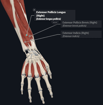
Originating from the middle third of the ulnar and interosseous membrane and inserting to the dorsal aspect of the distal phalanx of the first digit is the extensor pollicis longus. Extensor pollicis brevis originates from the middle half of the radius and interosseous membrane, inserting higher than the longus, via the base of the proximal phalanx of the first digit. The final muscle labelled in Figure 4 is the extensor indicis. Originating from the distal third of the ulnar and interosseous membrane and inserting onto the extensor expansion of the second digit. As their names suggest, these muscles extend the thumb and index fingers respectively.
The final muscle originating from the posterior forearm is the abductor pollicis longus. Originating from the proximal half of the ulnar, middle third of the radius, and interosseous membrane, inserting to the lateral aspect of the base of the first metacarpal bone. This insertion allows the abductor pollicis longus to both abduct and extend the thumb.
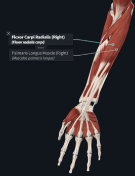
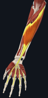
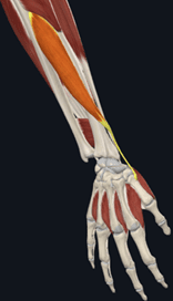
Figure 6 labels the flexor carpi radialis. Originating via the common flexor tendon of the medial epicondyle of the humerus, it then inserts to the palmar aspects of the bases of the second and third metacarpal bones, performing wrist flexion and abduction of the hand. Also labelled is the palmaris longus, with a shared origin of the flexor carpi radialis. The palmaris longus stabilises the palmar aponeurosis mentioned in the previous blog of the series, as it inserts to this structure and the flexor retinaculum of the hand.
The flexor digitorum superficialis is shown in figure 7 as a long, broad muscle of the anterior forearm, with a humeroulnar head, originating from the common flexor tendon and sublime tubercle of the ulna and ulnar collateral ligament, and a radial head via the proximal half of the anterior border of the radius. The tendons of these two heads then insert to the middle phalanges of the second to fifth digits, performing flexion of these digits.
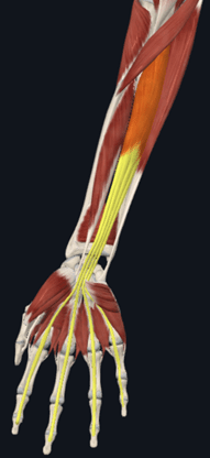
Deep to this lies flexor digitorum profundus with a lengthy origin from the coronoid process and the superior three quarters of the anteromedial aspect of the ulna. The flexor digitorum profundus inserts to the distal phalanges of the second to fifth digits, again, assisting in flexion of these digits.
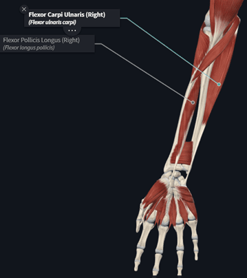
The last of the extrinsic muscles are the flexor carpi ulnaris and flexor pollicis longus, shown in figure 9. Flexor carpi ulnaris has a broad origin via the common flexor tendon, as well as olecranon and proximal two thirds of the ulna. It has an insertion to the carpals via the pisiform and hook of the hamate bone, as well as the base of the fifth metacarpal bone, allowing wrist flexion and adduction.
The flexor pollicis longus originates via the anterior surface of the radius and interosseous membrane, inserting to the base of the distal phalanx of the first digit, performing flexion of the thumb.
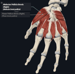
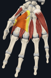
Remaining on the palmar aspect of the hand, the first intrinsic muscles are the abductor pollicis brevis and flexor pollicis brevis. These share a common insertion to the lateral aspect of the base of the proximal phalanx of the thumb. Their differing origins allow for the varied actions, with the abductor pollicis brevis originating from the tubercles of the scaphoid and trapezium bones and flexor retinaculum of the hand, and the flexor pollicis brevis originating from the trapezium, capitate and trapezoid bones more medially, as well as the flexor retinaculum of the hand.
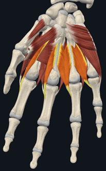
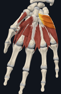
Figure 11 highlights the first to fourth lumbrical muscles of the hand. The origin of these muscles is the tendons of the flexor digitorum profundus, inserting to the extensor expansion of respective digits.
Figure 12 shows the palmaris brevis muscle, an important muscle to assist in hollowing the palmar surface of the hand for gripping. Originating from the palmar aponeurosis and flexor retinaculum of the hand, the palmaris brevis then inserts to the skin along the medial margin of the hand, over the thumb.
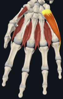
The adductor pollicis has an oblique and transverse head, both highlighted in figure 13. Both heads insert to the medial aspect of the base of the proximal phalanx of the thumb, but the oblique head originates more superiorly, via the capitate, and second and third metacarpal bones, whilst the transverse head originates from body of the third metacarpal bone.
Forming the medial margin of the hand is the abductor digiti minimi muscle. This originates from the pisiform bone and the tendon of the flexor carpi ulnaris, inserting to the medial aspect of the proximal phalanx of the fifth digit, allowing for abduction of the little finger.
Behind this, sits the flexor digiti minimi brevis muscle originating from the hook of the hamate bone and flexor retinaculum of the hand, sharing an insertion with the abductor digiti minimi, but performing flexion of the little finger.
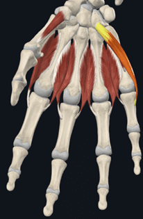
Highlighted in figure 16 are the oponens pollicis and oponens digiti minimi muscles. These originate from the tubercle of the trapezium and flexor retinaculum for the oponens policis, and the hook of the hamate bone and flexor retinaculum for the oponens digiti minimi. The two muscles insert to the body of the first metacarpal bones of the first and fifth digits respectively. These oponens pollicis allows the thumb to oppose toward the other fingers, and the oponens digiti minimi muscle opposes the little finger toward the thumb.
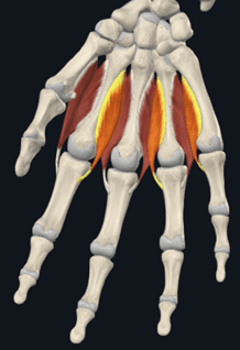
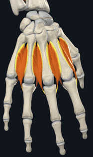
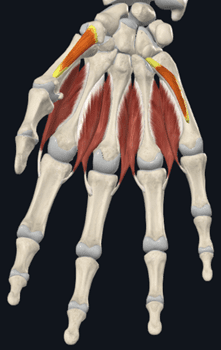
Finally, we have the first to third palmar interosseous muscles shown in figure 17, with the first to fourth dorsal interosseous muscles shown in figure 18.
The palmar interosseous muscles originate from the palmar aspect of the second, fourth, and fifth metacarpal bones, inserting to the extensor expansion of the second, fourth, and fifth proximal phalanx, adducting each of these digits.
The dorsal interosseous muscles originate from the medial and lateral aspects of the metacarpal bones they are located between, inserting to the extensor expansion of the second, third, and fourth proximal phalanx, opposing the action of the palmar interosseous muscles and abducting these digits.

Building our foundational knowledge of anatomy is a great way to gain confidence in the assessment and treatment of pathologies for the studied area. Familiarising yourself with the previous blogs in this series along with the muscular anatomy of the hand will assist in greatly increasing the specificity you can apply to your treatments, leading to greater outcomes for your patients.